FIG 2.
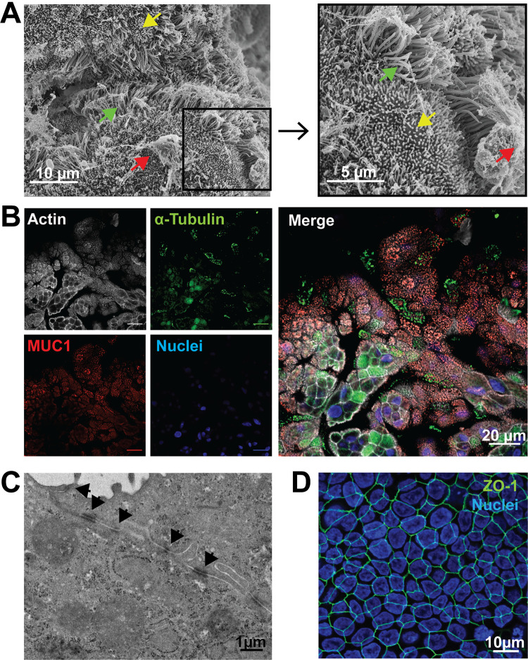
Primary human FTE cells cultured in an ALI form cilia, produce mucin-rich secretions, and form tight junctions. (A) Mucin, cilia, and microvilli were observed by scanning electron microscopy (SEM) on polarized FTE cells 24 days post-ALI. Arrows indicate mucin (red), microvilli (yellow), and cilia (green). (B) Mucin and structural protein of cilia were immunostained and observed by immunofluorescence in a single plane with individual channels (left) and a merged image (right) at 12 days post-ALI. Phalloidin for filamentous actin (white), α-tubulin for cilia (green), MUC1 for mucin (red), and DAPI (4′,6-diamidino-2-phenylindole; blue). Bars, 20 μm. (C) Cellular junctions, including tight junctions, were observed by transmission electron microscopy (TEM) of a cross section of an FTE cell junction 24 days post-ALI. Black arrows indicate cellular junctions. (D) Presence of the structural tight junction protein ZO-1 was observed by immunofluorescence at 5 days post-ALI (anti-ZO-1; green). Image contains DAPI staining by an extended focus view of the three-dimensional (3D) FTE cells.

