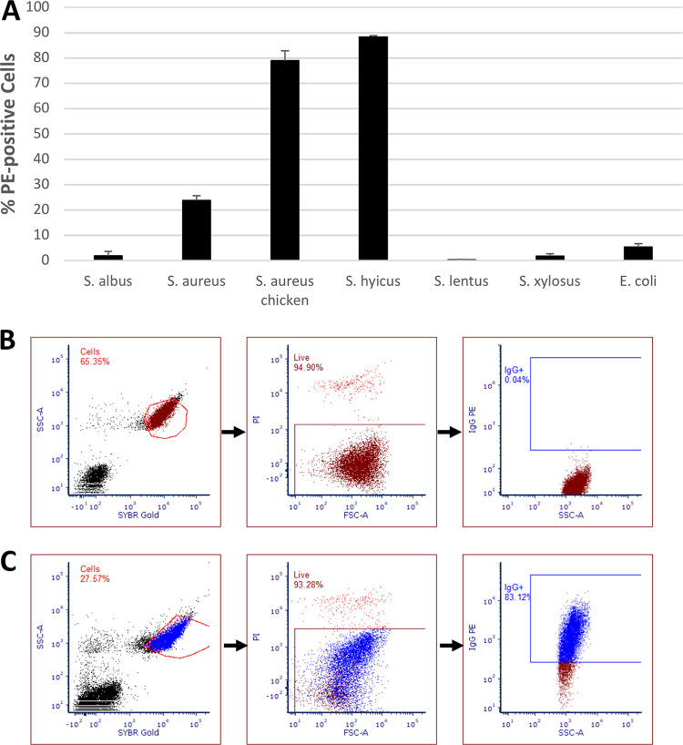
FIG 1.
(A) Mean percentages of cells of various Staphylococcus strains and E. coli binding to PE-conjugated mouse monoclonal anti-human CD44 antibody compared to unstained control S. aureus as measured using FCM (n = 2; bars show standard errors of the mean; P < 0.01, single-factor ANOVA). (B and C) Gating sequences for flow cytometric analysis of unstained S. aureus NCTC 8325 (B) and S. aureus chicken ready-meal isolate (C). Cells were distinguished from debris by using a plot of SYBR green fluorescence versus side-scattered light (SSC). Live cells were then distinguished from dead cells using a plot of forward-scattered light (FSC) versus PI fluorescence, and, finally, PE-positive cells were distinguished in a plot of SSC versus PE fluorescence, with the positive gate being drawn based on the fluorescence of unstained S. aureus cells.

