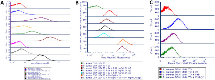
FIG 7.
(A to C) Overlays of histograms for the Alexa Fluor 647 fluorescence of S. aureus NCTC 8325, DSM 1104 and DSM 20231 incubated with mouse IgG, followed by staining with Alexa Fluor 647-conjugated goat anti-mouse IgG secondary antibody (A); S. aureus DSM 1104 incubated with various concentrations of purified goat IgG, followed by staining with mouse IgG and/or goat anti-mouse Alexa Fluor 647-conjugated secondary antibody (B); and S. aureus DSM 1104 cells incubated with whole, Fab, or F(ab′)2 fragment goat anti-mouse AF 647-conjugated antibodies (C). 2o, secondary antibody; Fab, Fab fragment goat anti-mouse IgG antibody; F(ab′)2, F(ab′)2 fragment goat anti-mouse antibody; Gt IgG, purified goat IgG; Mse IgG, mouse IgG; ITO, thiazole orange.

