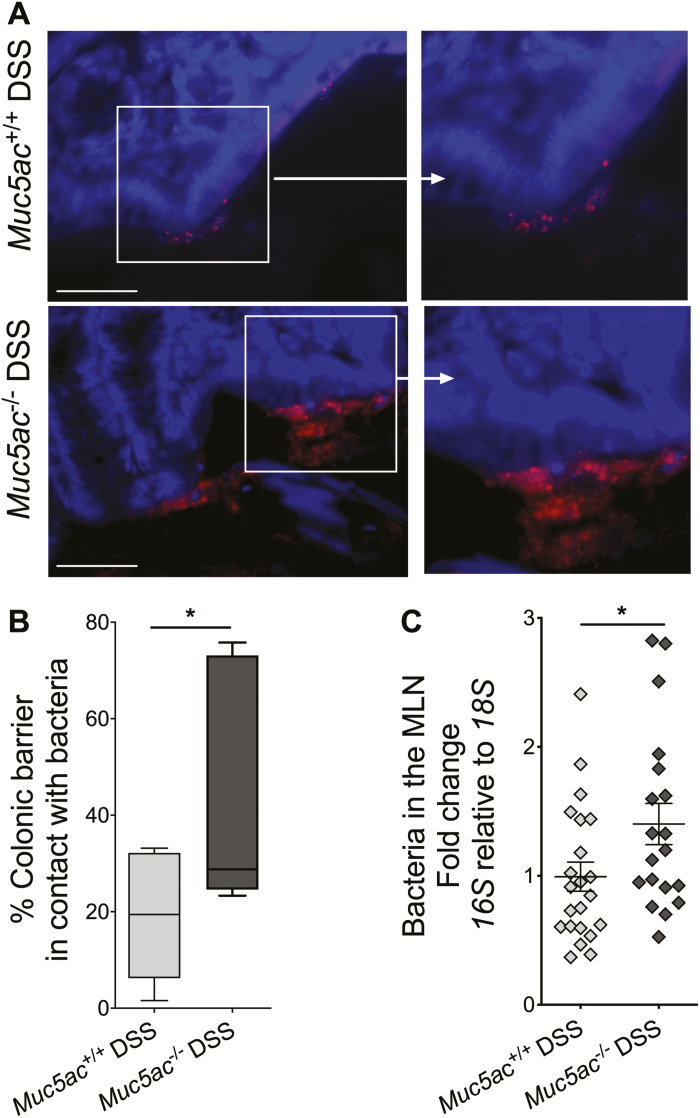
FIGURE 4.
Loss of Muc5ac increases bacterial contact with epithelium and translocation to MLN during experimental colitis. Administered DSS (3%) to Muc5ac-deficient mice (Muc5ac-/-) and their wildtype control mice (Muc5ac+/+) for 6 days, followed by water for 3 days. Whole colon and MLN were harvested. A, Performed 16S FISH to identify bacteria (red) in contact with the epithelium. Used 4′,6-diamidino-2-phenylindole as counterstain (blue). Bar represents 50 µm, images acquired at 40x. Images represent n = 7 to 9 mice/group. B, Length of epithelium in contact with bacteria displayed as percentage of total epithelial surface measured in 3 random images acquired from each mouse following 16S FISH as in Fig. 4A; n = 7 to 9 mice/group. C, Used 16S RT-PCR to detect bacterial load in MLN. Normalized PCR data to 18S; n = 19 to 22 mice/group. All results represent 3 independent studies and displayed as mean ± SEM. Two-tailed unpaired Student t test used to determine statistical differences. FISH indicates fluorescence in situ hybridization; SEM, standard error of the mean. *P< 0.05.

