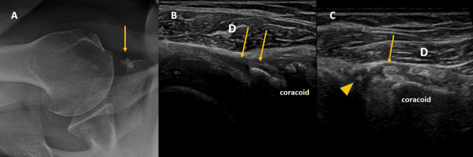
Fig. 4.
a Axial radiography showing calcifications medially to the coracoid process. b Axial ultrasound demonstrated that they were located at the coracoacromial ligament insertion (arrows). c Short axis ultrasound view of the PMi tendon (arrow) over the coracoid and medially to the coracoacromial ligament (arrowhead). d Deltoid

