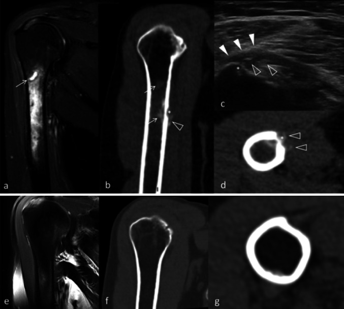
Fig. 3.
Deceptive aspect of calcification migration on MRI and diagnosis rectification by US. a MRI, STIR sequence, coronal view. Intra-medullary diffuse edema associated with small, liquid-like hyperintense foci of the bone marrow was secondarily identified as a calcification (arrow). b Non-contrast CT, coronal view. Cortical erosion (asterisk) with peripheral soft-tissues calcification (empty arrowhead) and intra-medullary calcifications (arrows). Notice the correspondence of the intra-medullary calcification with the MRI. c US, axial view. Typical aspect of calcification migration, with calcifications (empty arrowheads) sitting in the interstitial and deep fibers of the pectoralis major tendon (full arrowheads), in continuity with the cortical erosion (asterisk). No posterior acoustic shadowing is observed. d Non-contrast CT, axial view. Corresponding image, with humeral cortical erosion (asterisk) and calcifications on both sides (empty arrowheads). e, f, g Respectively, MRI, T2 Fat Sat sequence, coronal view; non-contrast CT, coronal view; non-contrast CT, axial view. Notice the restitutio “ad integrum” at 1-year follow-up. The intra-medullary edema, the cortical erosion, and the tendinous calcifications have entirely disappeared

