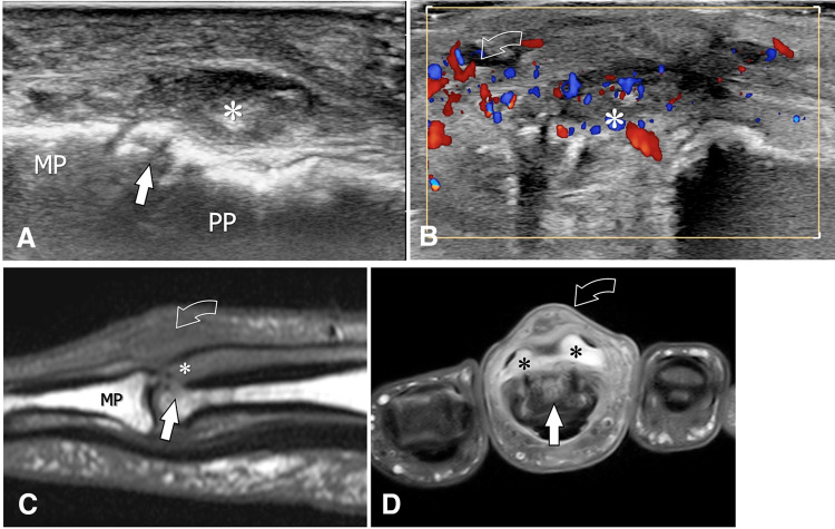
Fig. 13.
Postsurgical septic arthritis. Sagittal (a) and axial (b) colour Doppler sonograms, as well as sagittal T1-weighted (c) and transverse T1-weighted post-Gd-injection (d) MRI images, obtained at the proximal interphalangeal joint of the third finger in a patient presenting postsurgical septic arthritis. US shows hypertrophy of the synovium (asterisk) associated with erosion (arrow) of the dorsal aspect of the proximal phalanx (PP) head. In b, the important hypervascularisation of the synovium also extends into the periarticular soft tissues (curved arrow). MRI confirms the US appearance. In d, note the contrast enhancement of the synovium (asterisks), bone (arrow), and dorsal periarticular soft tissues (curved arrow)

