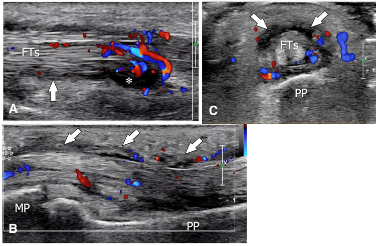
Fig. 14.
Postsurgical septic tenosynovitis. Sagittal (a, b) and axial (c) colour Doppler sonograms obtained over the flexor tendons (FTs) of the third finger in a patient presenting postsurgical septic tenosynovitis. US shows hypertrophy (arrows) and hypervascularisation of the common tendon sheath associated with a small amount of fluid (asterisk). MP middle phalanx, PP proximal phalanx

