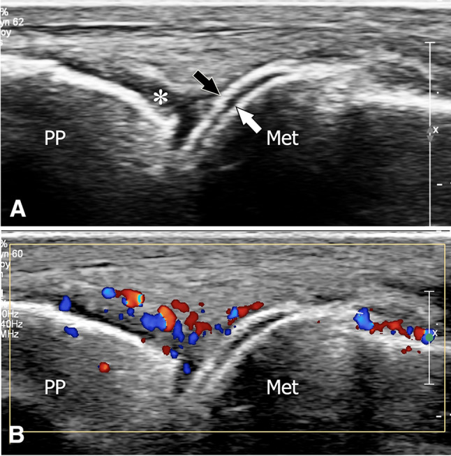
Fig. 16.

Acute postsurgical gout. Sagittal (a) and sagittal colour Doppler (b) sonograms obtained at the dorsum of the metacarpophalangeal joint of the index in a patient with gout presenting postsurgical monoarthritis. In a, note the hyperechoic linear band (black arrow) located at the surface of the cartilage (white arrow) of the metacarpal head. The hyperechoic line corresponds to the deposition of urate crystal on the cartilage surface. The asterisk points to synovial hypertrophy. In b, note the hypervascular changes inside the synovium. PP proximal phalanx, Met metacarpal
