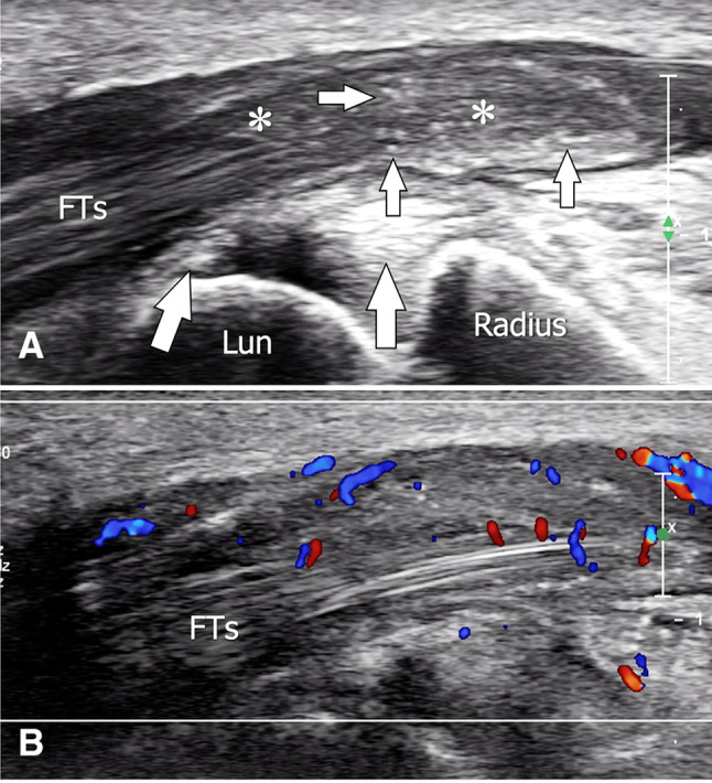
Fig. 17.

Acute postsurgical pseudogout. Sagittal (a) and sagittal colour Doppler (b) sonograms obtained over the carpal tunnel in a patient with crystal pyrophosphate deposition disease presenting postsurgical tenosynovitis of the flexor digitorum tendons (FTs). In a, note the hypertrophy of the synovial tendon sheath (asterisks) associated with multiple (small white arrows) internal hyperechoic spots due to crystal deposition. The larger arrows point to calcium deposition in the capsular palmar aspect of the radiocarpal joint. Lun lunate. In b, colour Doppler shows hypervascularity inside the inflamed tendon sheath
