
Fig. 19.
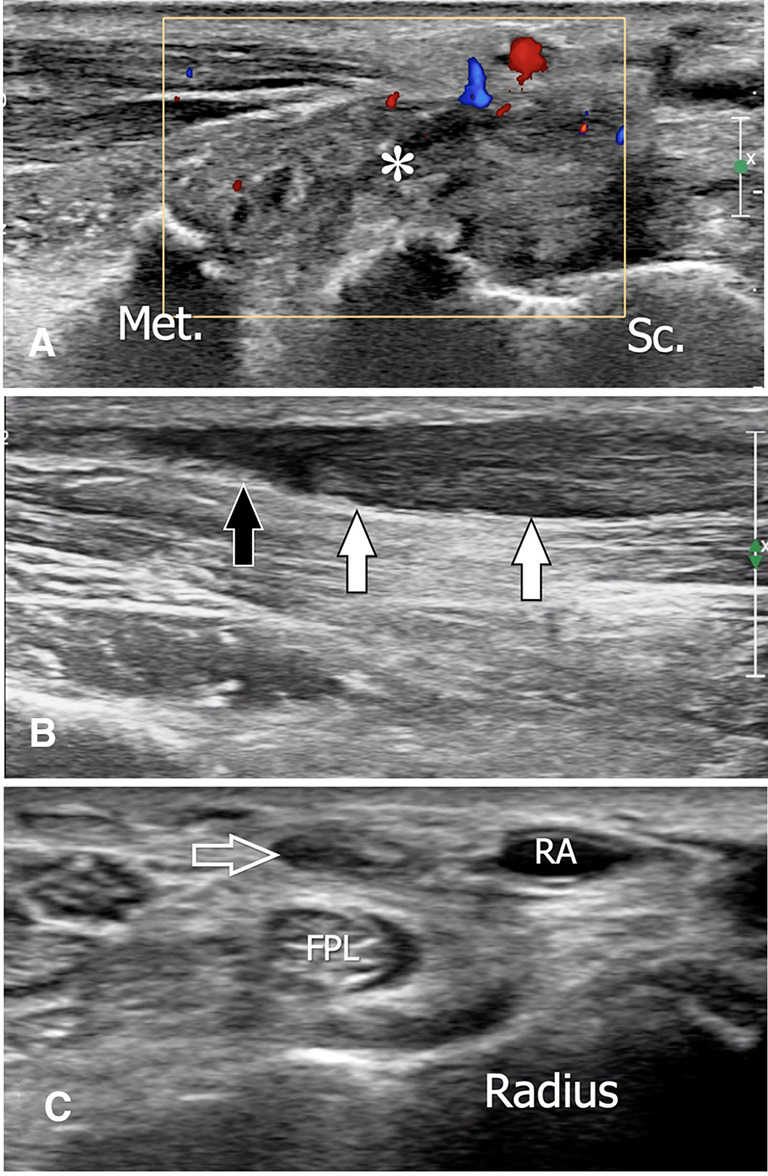
Tendon rupture after trapeziectomy. Sagittal (a, b) sonograms obtained over the wrist (a) and the distal metaphysis of the radius (b). Transverse (c) sonogram obtained over the distal epiphysis of the radius. The images were obtained for local pain in a patient with the previous trapeziectomy. In a, US shows local tear of the flexor carpi radialis tendon, which is replaced by a hypoechoic area (asterisk). b The tendon is retracted proximally and appears swollen and hypoechoic (white arrows). A small effusion is located inside the tendon sheath, distally to the tendon (black arrow). In c, the tendon sheath is empty (void arrow). Met first metacarpal, Sc scaphoid, FPL flexor pollicis longus, RA radial artery