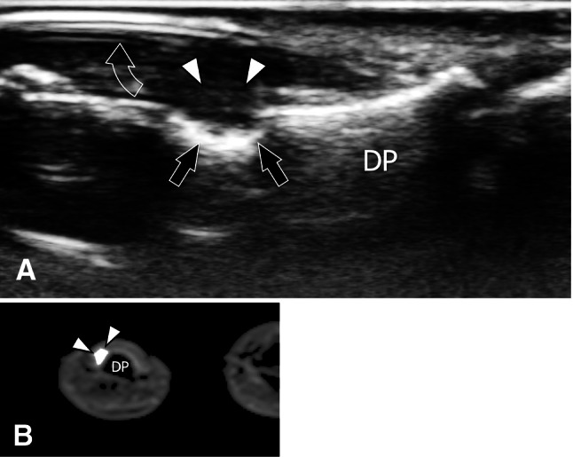
Fig. 29.

Persistent glomus tumour after negative surgical exploration. Sagittal sonogram (a) and transverse T1-weighted post-Gd-injection MRI image (b) obtained over the dorsal aspect of the distal phalanx (DP) in a patient presenting unchanged persistent excruciating pain after surgery for a glomus tumour. US shows the persistence of a glomus tumour (white arrowheads) located between the nail (void arrow) and the phalanx cortex. Note the surface erosion on the bone (black arrows) due to chronic local compression by the tumour. A second look at the MRI images revealed a small solid tumour showing contrast enhancement that was undetected at the first lecture
