
Fig. 30.
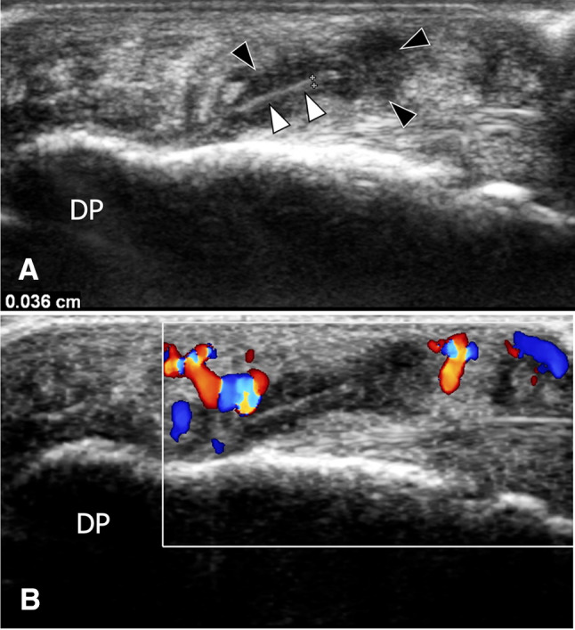
Persistent foreign body after negative surgical exploration. Sagittal greyscale (a) and colour Doppler (b) sonograms obtained on the fingertip of the fifth finger in a patient presenting persistent pain after negative surgical exploration for a suspected foreign body. In a, US shows a wood splinter (white arrowheads) located in the palmar soft tissue. The thickness (callipers) of the splinter is 0.36 mm. A hypoechoic area (black arrowheads) related to oedema surrounds the splinter. Colour Doppler shows local hypervascularisation related to inflammatory changes (b)