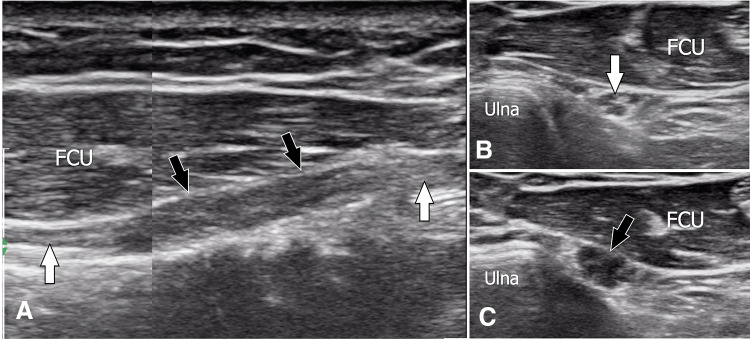
Fig. 4.
Posttraumatic in-continuity neuroma. Sagittal a and transverse b, c sonograms obtained over the dorsal sensitive branch of the ulnar nerve in a patient with previous local surgery and paraesthesia of the dorsum of the hand. In a, US shows a segmental enlargement of the nerve (black arrowheads), which appears hypoechoic. Note the loss of the normal internal fascicular structure. The appearance is typical of posttraumatic in-continuity neuroma. The white arrows point to the normal proximal and distal nerves. Axial US obtained proximal to the neuroma (b) depicts a normal nerve. Pathologic nerve changes are confirmed at the axial sonograms obtained over the neuroma (c). FCU flexor carpi ulnaris

