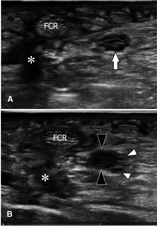
Fig. 5.

Posttraumatic in-continuity neuroma complicating osteosynthesis of the distal radius. Axial sonogram (a) obtained from the proximal shows the normal appearance of the median nerve (arrow). More distally, the nerve shows an in-continuity neuroma as an area of loss of fascicular structure (black arrowheads) located at its radial portion. Note the disappearance of the normal fascicular pattern inside the neuroma, while the fascicles located in the ulnar portion of the nerve (arrowheads) are normal. The appearance is evocative of a nerve trauma due to surgical distractors. The asterisks point to a normal irregularity of local soft tissues related to surgery
