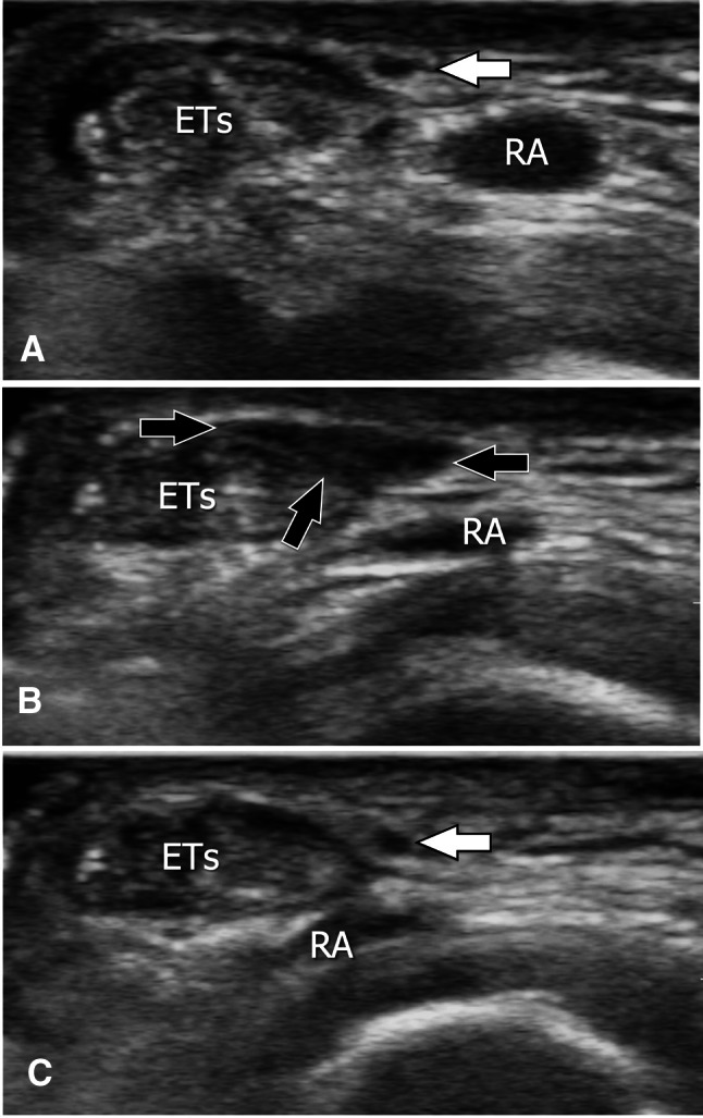
Fig. 6.

Axial sonograms (a–c) obtained from proximal (a) to distal (c) over the superficial branch of the radial nerve in a patient with previous surgical treatment for De Quervain tenosynovitis. In a, the nerve appears normal (white arrow). More distally (b), the nerve is not detectable because it runs inside a hypoechoic area (black arrows) related to postsurgical fibrosis. Further distally (c), note the normal appearance of the nerve (white arrow) surrounded by normal soft tissues. ETs extensor tendons of the first compartment, RA radial artery
