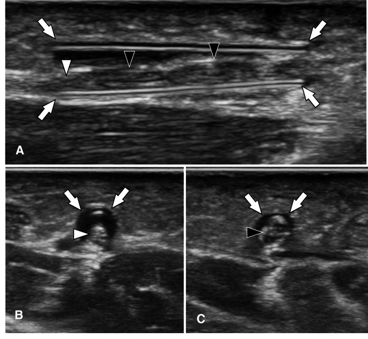
Fig. 7.
US appearance of nerve conduits. Longitudinal (a) and axial (b, c) sonograms obtained over a nerve conduit located at the palmar-radial aspect of the distal forearm. The nerve conduit presents at US as a tubuliform structure with hyperechoic walls. The nerve located inside the conduit appears normal (white arrowheads) proximally. Note the thickening and hypoechogenicity of the nerve (black arrowheads) in the middle of the conduit

