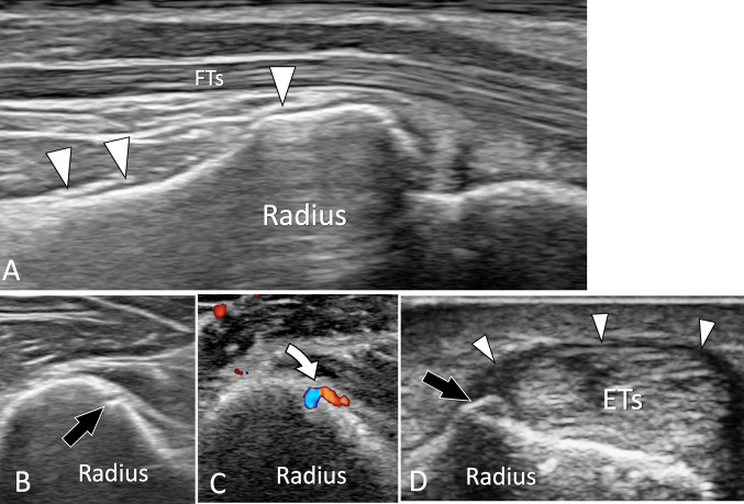
Fig. 1.
US of normal adult bone. a Longitudinal sonogram obtained aver the palmar aspect of the distal radius. The normal periosteum of the radius (white arrowheads) appears as a thin hypoechoic band located on the bone palmar cortex. b, c Axial conventional (b) and colour Doppler (c) sonograms obtained at the middle of the radius. In b note, a focal break in the cortex which corresponds in c to the location where the nutrient artery (curved arrow) enters the bone. d Axial US image obtained over the radial aspect of the distal radius. US shows a small bone ridge at the insertion of the retinaculum (white arrowheads) of the first compartment of the extensor tendons (ETs)

