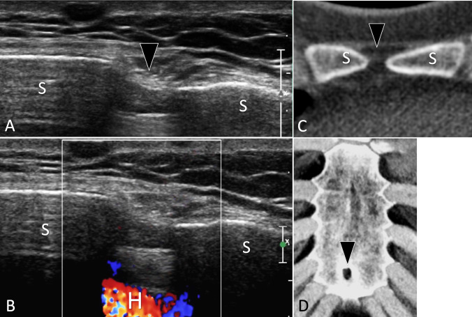
Fig. 10.
Bone hypoplasia. Sternal foramen. a, b Axial grey scale (a) and colour Doppler (b) US of the anterior cortex of the sternum. c, d Axial native (c) and d 3D reconstructed CT of the sternum. In a. US shows a sternal foramen as a focal discontinuity (black arrowhead) of the anterior cortex of the sternum (S). Note the regular cortex surrounding the foramen, absence of periosteal reaction, or soft-tissue masses. In b, note visualisation of the heart (H) due to a full-thickness interruption of the sternum. c, d CT confirms the diagnosis of a sternal foramen

