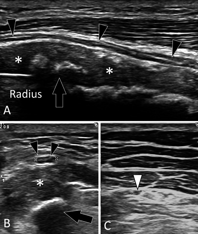
Fig. 17.

Complications of fractures. Nerves lesions. a, c Sagittal (a) and axial (b, c) sonograms in a patient with the previous fracture of the radius shaft. In a the superficial branch of the radial nerve (black arrowheads) is hypoechoic and thickened. The nerve is displaced by bone callus (asterisks) which is partially calcified. In b, note the swelling (black arrowheads 3.5 mm) of the nerve in the axial plane compared with the nerve (white arrowhead 1.6 mm) imaged at a more distal level (c)
