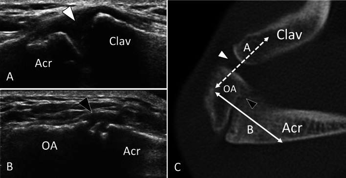
Fig. 2.
Non-fusion of ossification centres. Asymptomatic os acromiale. US (a, b) with axial CT (c) correlation in a patient with os acromiale. Sonogram a obtained over the acromioclavicular joint (line A in C) shows the normal joint (white arrowhead). Note smooth borders of bone ends. b US image obtained over the Os acromiale (OA) (line B in C) exhibits a focal interruption (black arrowhead) of the hyperechoic line corresponding to the bone surface due to non-fusion of the os acromiale. In b, c, note the rough borders of the non-fusion

