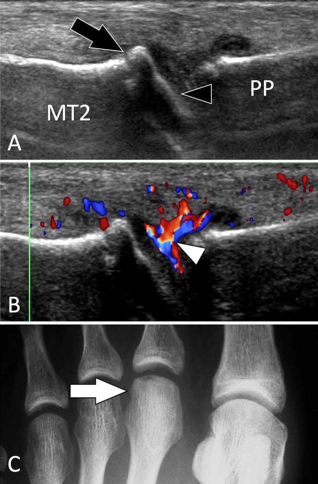
Fig. 22.

Intra-articular fractures. Freiberg disease. Sagittal conventional (a) and sagittal colour Doppler (b) sonograms on the dorsal aspect of the second metatarsophalangeal joint. c A-P radiograph. Sonograms illustrate a flattened joint surface (black arrowhead) associated with a small dorsal osteophyte of the second metatarsal (MT2). In b, note synovial hypertrophy with hyperaemia (white arrowhead). Radiograph shows a dense head of the metatarsal with a slightly fat articular surface
