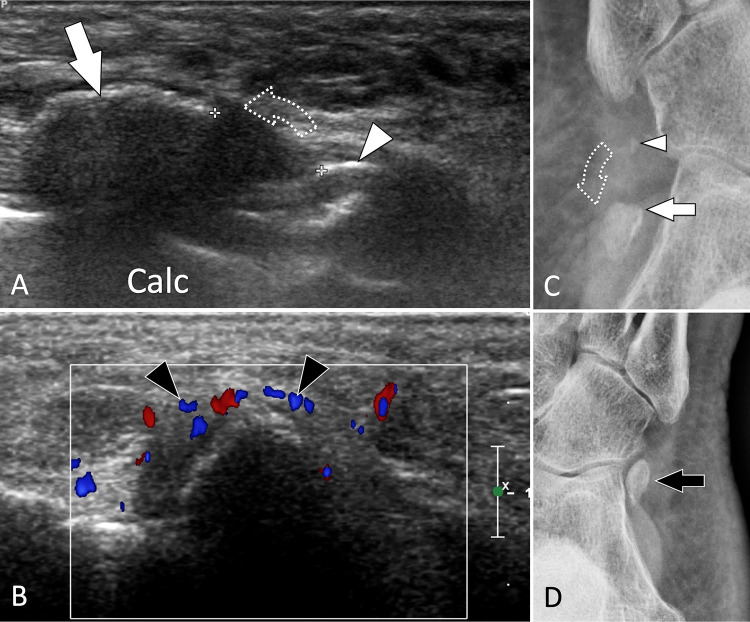
Fig. 28.
Bone avulsion. Os peroneum. a, b Axial oblique conventional (a) and coronal oblique colour Doppler (b) sonograms obtained on the Os peroneum in a patient with recent trauma. Images show a fracture of the ossicle with the presence of a proximal large (white arrow) and a small distal (white arrowhead) fragment. Note proximal displacement (dotted arrow) of the proximal fragment due to traction through the peroneus longus tendon. b Colour Doppler shows local hyperaemia (black arrowheads). c, d Internal oblique radiographs of the affected (c) and contralateral (d) foot. In c, note the fracture avulsion of the upper pole of the os peroneum associated with proximal displacement confirming the US appearance. d A normal os peroneum

