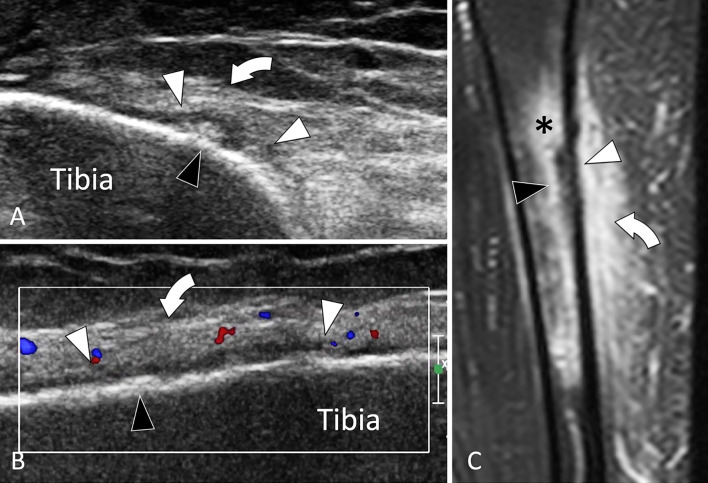
Fig. 31.
Stress fracture. Tibial shaft. a, b Axial (a) and coronal oblique (b) sonograms, d Coronal t2-weighted fat sat MR image. Sonograms show a focal irregularity of the bone surface (black arrowheads) associated with periosteal thickening (white arrowheads) and oedema (curved arrows) of the subcutaneous soft tissues. c MR illustrates in a better detail oedema (asterisk) of soft tissues but also bone marrow oedema

