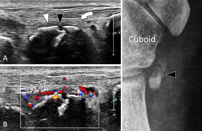
Fig. 33.
Stress fracture. Os peroneum. a, b Axial oblique conventional (a) and colour Doppler (b) sonograms. c Internal oblique radiograph centred at the cuboid region. In a, b US shows discontinuity (black arrowhead) of the cortex of the os peroneum associated with mild periosteal thickening (white arrowhead) and associated oedema (curved arrow) of the adjacent soft tissues. The inflamed periosteum shows important hypervascular changes (void arrowhead) in b. Note associated oedema (curved arrows) of the subcutaneous soft tissues. In c, the ossicle, enlarged and irregular, presents a fracture with mild displacement. MRI (not shown) confirmed the US data showing bone marrow oedema of the ossicle and associated soft-tissue hyperaemia

