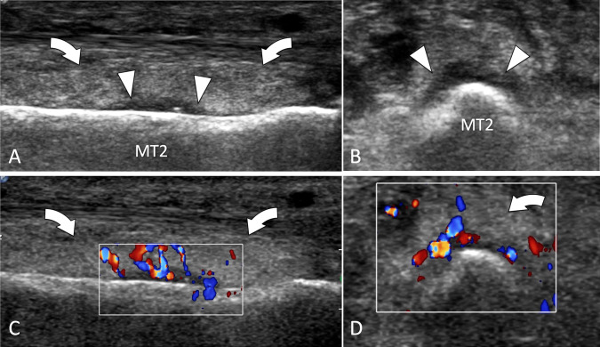
Fig. 34.
Stress fracture. Metatarsal. a, d Axial and sagittal conventional (a, b) and colour Doppler (c, d) sonograms. US shows periosteal thickening (white arrowheads) of the second metatarsal (MT2). The inflamed periosteum shows important hypervascular changes in the colour Doppler images (c, d). Note associated oedema (curved arrows) of the subcutaneous soft tissues

