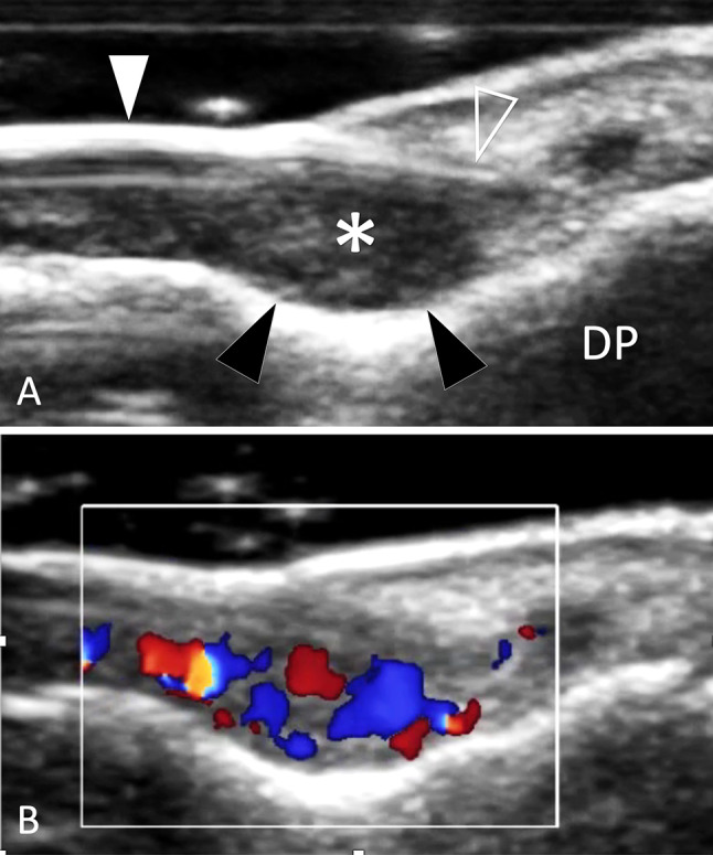
Fig. 40.

Tumour. Glomus tumour. a, b Sagittal conventional (a) and colour Doppler (b) sonograms obtained over distal phalanx (DP) of the third finger. The glomus tumour (asterisk) presents as an ill-defined ovular mass located between the nail (white arrowhead) matrix (void arrowhead) and the superficial bone cortex. Note scalloping of the cortex (black arrowheads) due to chronic local pressure. In b, the tumour shows internal vascular signals
