
Fig. 5.
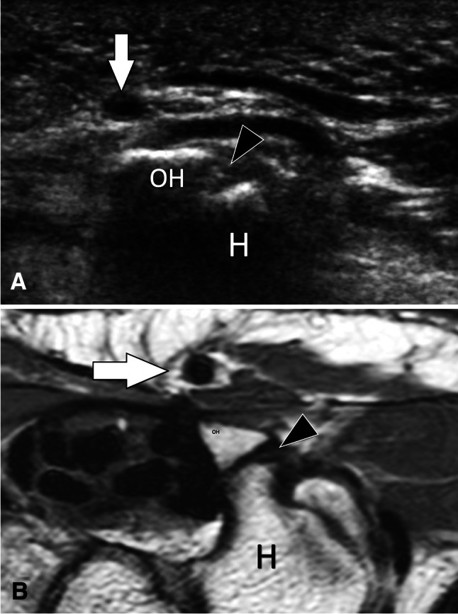
Non-fusion of ossification centres. Asymptomatic os hamuli proprium. US (a) with axial T1-weighted MR image (b) correlation in a patient with os hamuli proprium. Sonogram a obtained over the palmar aspect of the wrist shows a focal interruption (black arrowhead) of the hyperechoic line of the ulnar aspect of the hamatum (H) corresponding to non-fusion of the ossification centre. In B, the MR image confirms the US findings. White arrows = ulnar artery