Fig. 6.
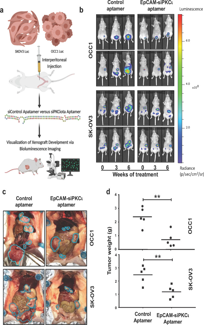
EpCAM aptamer-delivered PKCι siRNA suppresses intraperitoneal xenograft development. a Scheme of experimental procedure. Luciferase-expressing SK-OV-3 or OCC1 cells (107 cells/mouse) were intraperitoneally inject into athymic nude mice. Once tumors were detected, mice were divided into two groups: one was treated with control aptamer and the other with EpCAM-siPKCι aptamer. Xenograft development was monitored using the IVIS bioluminescence imaging system. b Images of the xenograft tumors using the Xenogen IVIS at point of treatment (marked 0), 3 weeks and 6 weeks of treatment. Two representative mice from each group were included. The image data is displayed in radiance or photons/sec/cm2/steradian. c Mice were sacrificed after 7 weeks of treatment and images were taken after dissection. Tumor are circled in teal. d Tumor implants were collected and weighed. Average of tumor weights for each group were calculated (n = 5). ** indicates P < 0.01 EpCAM-siPKCι aptamer versus control determined by ANOVA and student t tests

