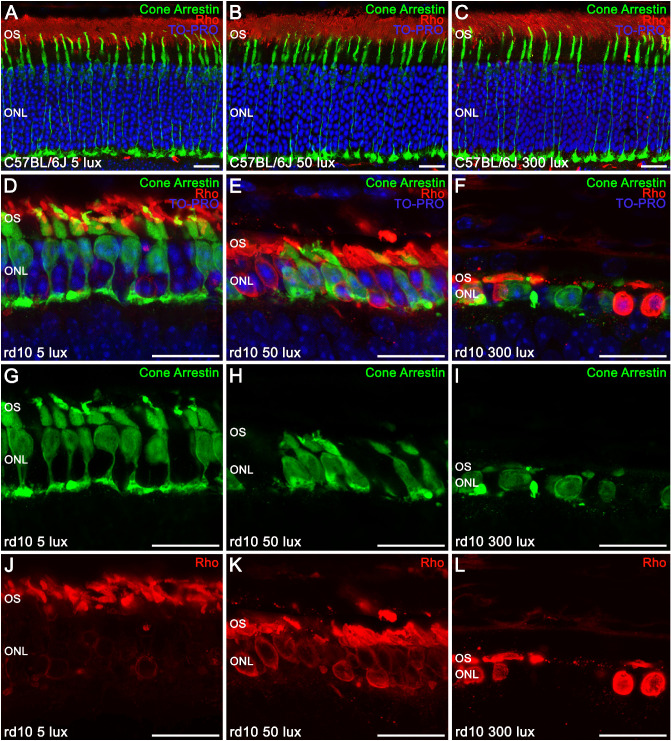
Figure 5.
Effect of environmental light conditions on photoreceptor morphology. (A–L) Cross-sectional cryosections of retinas from P30 C57BL/6J mice (A–C) and rd10 mice (D–L) reared under 5, 50, and 300 lux cyclic light and immunolabeled against cone arrestin (cone cells, green), rhodopsin (Rho, rod cells, red), and TO-PRO 3 (nuclei, blue). In C57BL/6J mice, merged images showed a normal cone photoreceptor morphology and rhodopsin distribution (A–C). In rd10 mice, merged images (D–F) showed a light-dependent degeneration of the photoreceptors, with an abnormal distribution of rhodopsin (G–I) accumulated in the rod cell body and a progressive morphological alteration of the cones (J–L). OS, outer segments; ONL, outer nuclear layer. Scale bars: 20 µm.

