
Fig. 5. Actin patches can be imaged using LIVE-PAINT in live yeast.
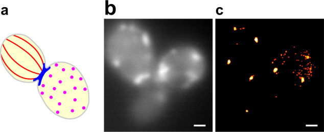
a Cartoon showing the three distinctive actin structures that have been observed in fixed and immunostained S cerevisiae: actin cables (red), actin rings (blue), and actin patches (magenta). b Diffraction limited image of SYNZIP18-Act1p + SYNZIP17-mNG. c LIVE-PAINT super-resolution image constructed from 200 s video imaging SYNZIP18-Act1p + SYNZIP17-mNG (50 ms exposure per frame and a laser power density of 3.1 W/cm2). Number of localization events obtained: 778. Only localization events with precision <30 nm were used to construct the super-resolution image. Scale bars are 1 μm.