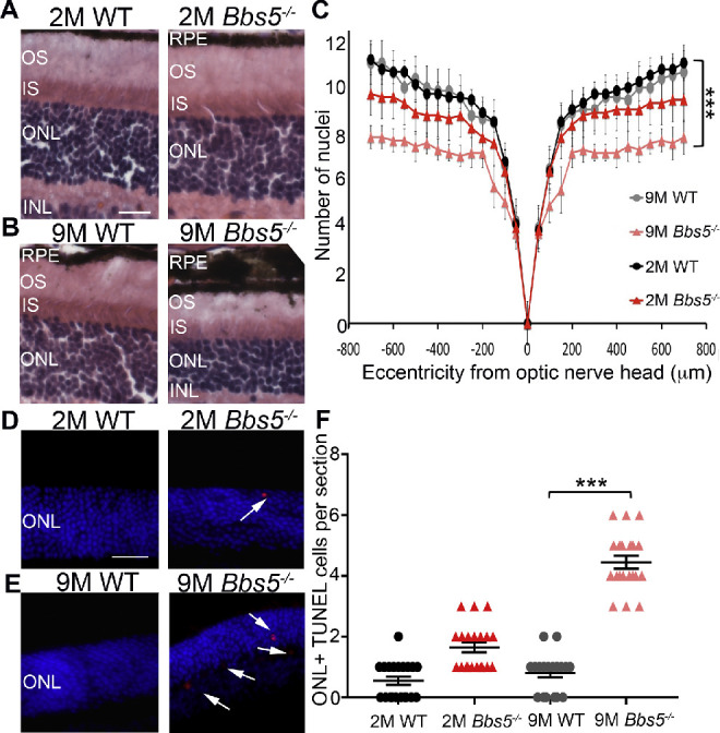
Figure 2.

Congenital absence of BBS5 results in retinal degeneration. (A, B) H&E staining of retina sections from WT and Bbs5–/– 2-month-old mice (2M) and 9-month-old mice (9M). Retina layers are indicated as follows: retinal pigment epithelium (RPE), outer segment (OS), inner segment (IS), outer nuclear layer (ONL), and inner nuclear layer (INL). Scale bar: 50 µm. (C) Morphometric analysis of nuclei counts at different distances from the optic nerve head using Student's t-test revealed only slight retinal degeneration in 2M Bbs5–/– compared to statistically significant retinal degeneration in 9M Bbs5–/–. (D, E) TUNEL staining images for apoptosis (red) in 2M and 9M retinas. DAPI stained nuclei are blue. Scale bar: 50 µm. (F) Graph of TUNEL quantification in 2M and 9M retinas. Kruskal–Wallis nonparametric ANOVA with post hoc Mann–Whitney comparisons yielded ***P < 0.05 (n = 6 per group; mean ± SEM).
