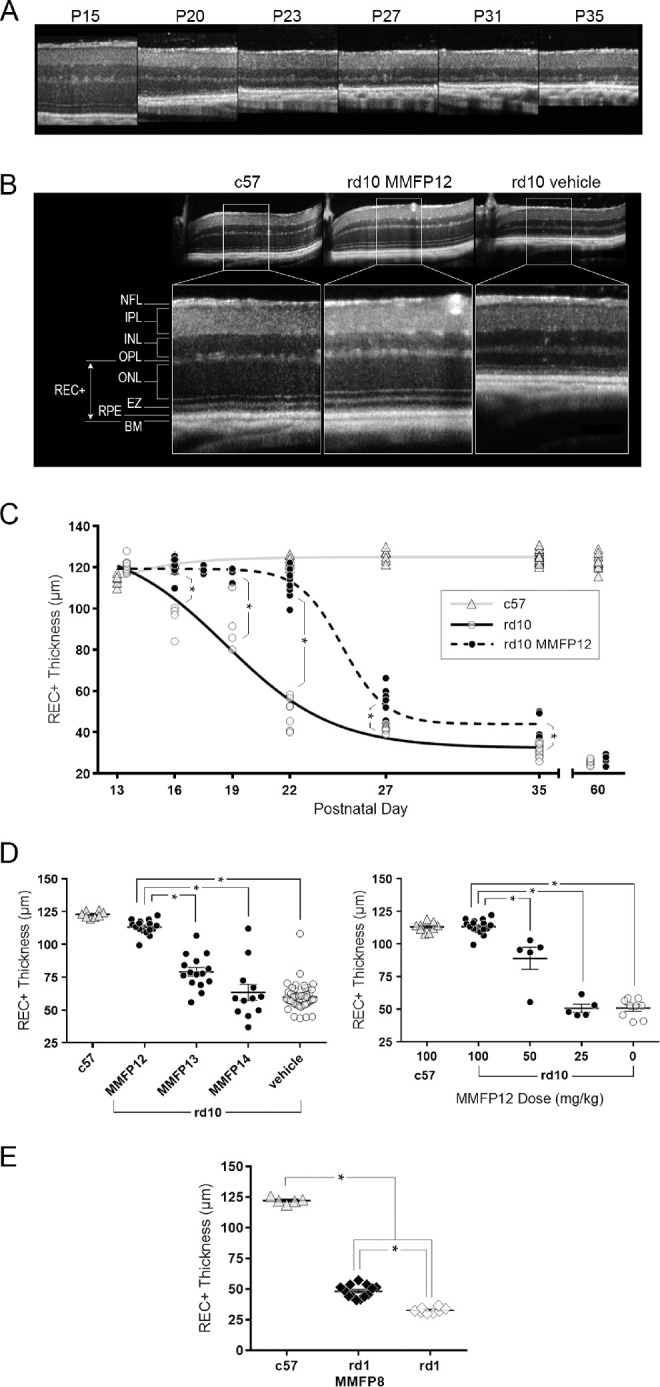
Figure 1.

Effect of MMF on the outer retina in rd10 and rd1 mice. (A) OCT images of naïve rd10 mice degeneration over time from P15 to P35, which shows considerable loss in the outer retina by P23. (B) Representative OCT images of the retina at P22 showing that rd10 mice treated with MMF 100 mg/kg starting at P12 (MMFP12) had similar outer retinal structure to c57 mice, as compared with the degeneration seen in rd10 mice treated with vehicle. (C) REC+ thickness from rd10 mice treated with MMFP12 were significantly greater than naïve rd10 mice up to P35 even though the benefit declined after P22: P16 (n = 8 and 4, P < 0.0001), P19 (n = 6 and 4, P <0.0001), P22 (n = 16 and 9, P < 0.0001), P27 (n = 8 and 7, P = 0.0021), P35 (n = 4 and 12, P = 0.0029), and P60 (n = 8 and 8, P > 0.99). The REC+ thickness in c57 mice was stable across time. (D) MMF treatment of rd10 mice starting at P12 (n = 16) produced greater REC+ protection by P22 as compared with initiating treatment at P13 (MMFP13, n = 15, P < 0.0001) or P14 (MMFP14, n = 12, P < 0.0001) as well as vehicle-treated rd10 mice (n = 49, P < 0.0001). The dose response of MMFP12 treatment in rd10 mice showed that 100 mg/kg (n = 16) produced significantly better efficacy compared with 50 mg/kg (n = 5, P < 0.0001), 25 mg/kg (n = 5, P < 0.0001), and no treatment (n = 9, P < 0.0001). (E) The rd1 mice treated with 100 mg/kg MMF initiated at P8 (MMFP8) had significantly thicker REC+ than naïve rd1 mice at P15 (n = 13 and 8, P < 0.0001), albeit the effect was more modest than in rd10 mice. INL, inner nuclear layer; EZ, ellipsoid zone; BM, Bruch's membrane; *P ≤ 0.05.
