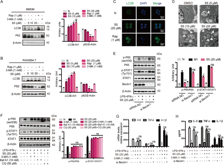
Fig. 4. S5 increased macrophage autophagy in vitro.
a, b BMDMs and RAW264.7 cells were treated with 5, 10, and 20 μM of S5 and 1 μM of rapamycin, 1 mM 3-MA or the same volume of DMSO, respectively, for 6 h. S5 promoted autophagy by enhancing LC3B-II/I ratio and decreasing P62 expression. Data are means ± SEM of five independent experiments. *P < 0.05, **P < 0.01 vs. DMSO group. c LC3B expression in RAW264.7 cells were analyzed by immunofluorescence staining. Scale bar, 20 μm. d BMDMs were fixed and examined for autophagosome formation by transmission electron microscopy. Scale bar, 0.5 μm. e, f The inhibition effect of S5 on M1 polarization signal pathway was repressed with Beclin1 knockdown (e) or autophagic inhibitor 3-MA, BafA1, and CQ (f). Data are means ± SEM of five independent experiments. ns nonsignificant, *P < 0.05, **P < 0.01 vs. LPS and IFN-γ group. g, h Il-6, TNF-α, and Il-1β mRNA expression were detected by RT-PCR (g), and protein level in medium was detected by ELISA assay (h). Data are means ± SEM of five independent experiments. ns nonsignificant, *P < 0.05, **P < 0.01, ***P < 0.001 vs. LPS and IFN-γ group.

