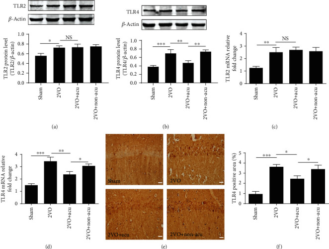
Figure 2.

Acupuncture inhibits the expression of TLR4, but not TLR2, in the hippocampus of 2VO rats. (a, b) Representative Western blots and the densitometric analysis of TLR2, TLR4, and its corresponding β-actin bands in protein levels were determined at day 14 after acupuncture treatment using Western blot analysis. The lanes (from left to right) represent the sham, 2VO, 2VO+Acu, and 2VO+Non-acu groups (n = 6). (c, d) The mRNA expressions of TLR2 and TLR4 in the hippocampus were assessed by quantitative real-time PCR at day 14 after acupuncture treatment in the sham, 2VO, 2VO+Acu, 2VO+Non-acu groups (n = 6). (e) Representative photomicrographs of TLR4 immunohistochemistry are shown. (f) Graphic presentations show the positive area of TLR4 in the hippocampal CA1 region at day 14 after acupuncture treatment (n = 6). ∗P < 0.05, ∗∗P < 0.01, and ∗∗∗P < 0.001, compared as indicated.
