
Figure 3.
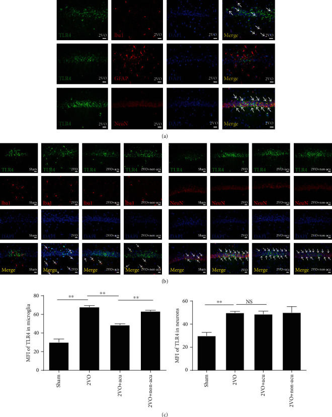
Acupuncture reduces the expression of TLR4 in microglia. (a) Representative photomicrographs of TLR4 (green) colocalized with neurons (NeuN, red), microglia (Iba1, red), and astroglia (GFAP, red) in the hippocampus of 2VO rats are shown. Nuclei were stained with DAPI (blue). (b) Representative photomicrographs of TLR4 immunofluorescence are shown for each condition. TLR4 localizes to the respective cellular marker with areas of overlap appearing yellow in the merged image (white arrow). Scale bars: 50 μm. (c) The graph shows the MFI of TLR4 in microglia or neurons in the hippocampus of 2VO rats (n = 6). ∗∗P < 0.01, compared as indicated. MFI: mean fluorescence intensity; NS: no significance.