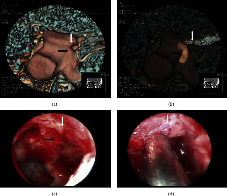
Figure 1.

A 42-year-old man with traumatic optic neuropathy. The VR image and endoscopic image of optic nerve and internal carotid artery present on the inner wall of sphenoid sinus. White arrow shows optic canal; black arrow shows internal carotid artery. (a) The VR image of the medial wall of the sphenoid sinus. (b) The fuse VR image of the medial wall of the sphenoid sinus, the optic nerve, and the internal carotid artery. (c) The anatomical structure of the medial wall of the sphenoid sinus under endoscopy. (d) The optic nerve under endoscopy.
