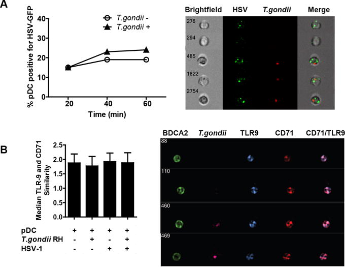
Figure 3. T. gondii infection neither affects the uptake of HSV-1 nor the subcellular localization of TLR9.

A, Purified pDC were infected with T. gondii-mCherry-RH at an MOI of 4 for 1 hr, then incubated with HSV expressing GFP capsid at MOI of 10. At indicated times, cells were extensively washed with ice cold PBS EDTA to remove surface bound virus and fixed with PFA. Samples were collected with the Amnis ImageStream and percent of pDC positive for HSV-GFP was determined using IDEAS analysis software and compared between T. gondii uninfected and infected pDC. On the right are the representative images of T. gondii uninfected and infected HSV-GFP positive pDC and on the left is a time-course analysis of HSV-GFP uptake in the presence or absence of T. gondii. B, Amnis ImageStream was used to determine the similarity score between TLR9 and recycling endosome marker CD71 following 30 min exposure to HSV. Mean similarity scores ± SEM from three independent experiments are shown. On the right, representative images of four cells showing TLR9 distribution in CD71-positive compartments are shown.
