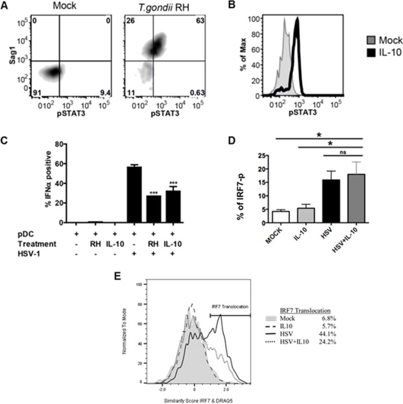
Figure 5. T. gondii, like IL-10 inhibits STAT3 phosphorylation and IFN-α production through blockade of IRF-7 translocation but not phosphorylation.

A, Phosphorylation of STAT3 (pY705) was measured in T. gondii-infected pDC 5-hr post- infection using intracellular phospho-flow cytometry. T. gondii infection of pDC (identified by HLA-DR-Pac-blue and CD123-APC) was assessed using intracellular staining against Sag1. Data are representative of three independent experiments. B, PBMC were treated with IL-10 (50 ng/ml) for 15 minutes. Data shown are histogram overlays of IL-10-induced phosphorylation of STAT3 in pDC from PBMC population. Data are representative of three independent experiments. C, Summary figure for IL-10 and T. gondii inhibition of IFN-α production as compared to HSV stimulation of pDC using intracellular flow cytometry as described in A, B (n=3, *** p<0.01 vs. HSV-stimulated). D, PBMC were pretreated with or without IL-10 (50 ng/ml) for 1hr, then the cells were stimulated with HSV-1 (MOI=1) for 3hrs. Phospho-flow analysis was performed for IRF-7 activation in pDC as described in Figure 4A. Data represent the mean ± SEM of three independent experiments; E, IL-10 mediated inhibition of HSV-induced IRF7 nuclear translocation. Enriched pDC were pretreated with or without IL-10 (50 ng/ml) for 1hr, then the cells were stimulated with HSV-1 for 5hrs. IL-10 mediated inhibition of HSV-induced IRF7 nuclear translocation was quantified using Amnis ImageStream as described for Figure 4. Data are representative of three independent experiments.
