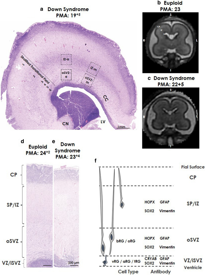
Fig. 1.
Coronal section of regions of interest. Photomicrographs of Haemotoxlin and Eosin (H&E) stained (a) coronal section from the frontal lobe of a Down syndrome case at 19+5 weeks PMA showing the stratified transitional field (1–6) and regions of interest. b, c Coronal T2-weighted MRI images from (b) control, 23 weeks PMA and (c) Down syndrome, 22+5 weeks PMA, highlighting different regions based on T2-weighted signal intensities. Higher magnification of H&E stained frontal lobe coronal sections from (d) age-matched Euploid case at 24+2 weeks PMA, and (e) Down syndrome case at 23+4 weeks PMA. F Schematic of regions of interest and associated protein labels of radial glia. Scale bars indicated (a) 1 mm, (b, c) 200 µm. Abbreviations: –a anterior, CN caudate nucleus, CC corpus callosum, CP cortical plate, IZ intermediate zone, LV lateral ventricle, -m medial, RG radial glia (aRG; apical, bRG basal, oRG outer, vRG; ventral, tRG; truncated), SVZ subventricular zone (iSVZ; inner, oSVZ; outer), PMA post-menstrual age, STF stratified transitional field, SP subplate, VZ ventricular zone

