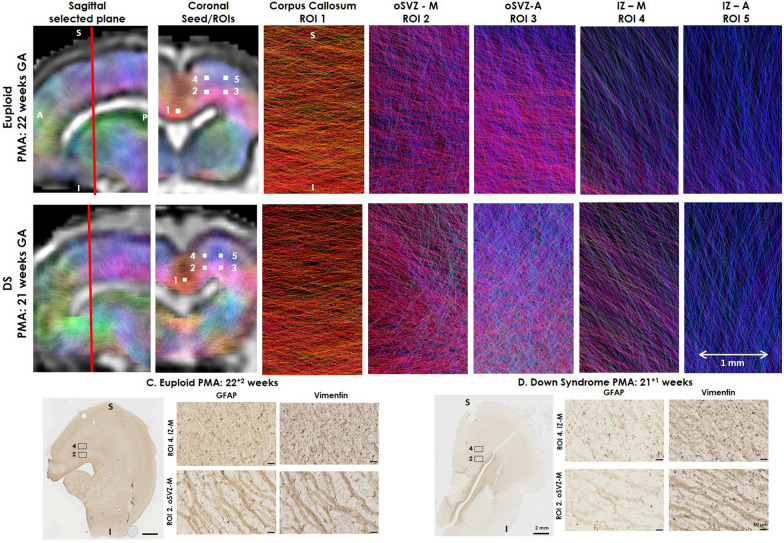
Fig. 6.
Fetal diffusion MRI and immunohistochemistry in the fetal brain. Upper panels show a (A) Euploid (22 GW) and (B) DS (21 GW) fetal brain, following in vivo fetal MRI in sagittal plane, and 5 seed/regions of interest (ROI), approximately 1 × 1 × 1 voxel, (2 mm), corresponding to immunohistological ROIs assessed and the corpus callosum. The colour-coding of the tractography is based on the standard red, green, blue code to indicate directionality; red for left–right, green for anterior–posterior and blue for dorsal–ventral. The lower panel reflect GFAP and Vimentin labelled radial glia, with images rotated in a comparable plane to the fetal MRI images in the (C) Euploid and (D) DS brain, showing direction of fibres in comparable ROI 2 (oSVZ-M) and ROI 4 (IZ-M) stained with GFAP and Vimentin. S superior, I inferior. Scale bar indicates 2 mm on low magnification and 50 µm on high magnification photomicrographs

