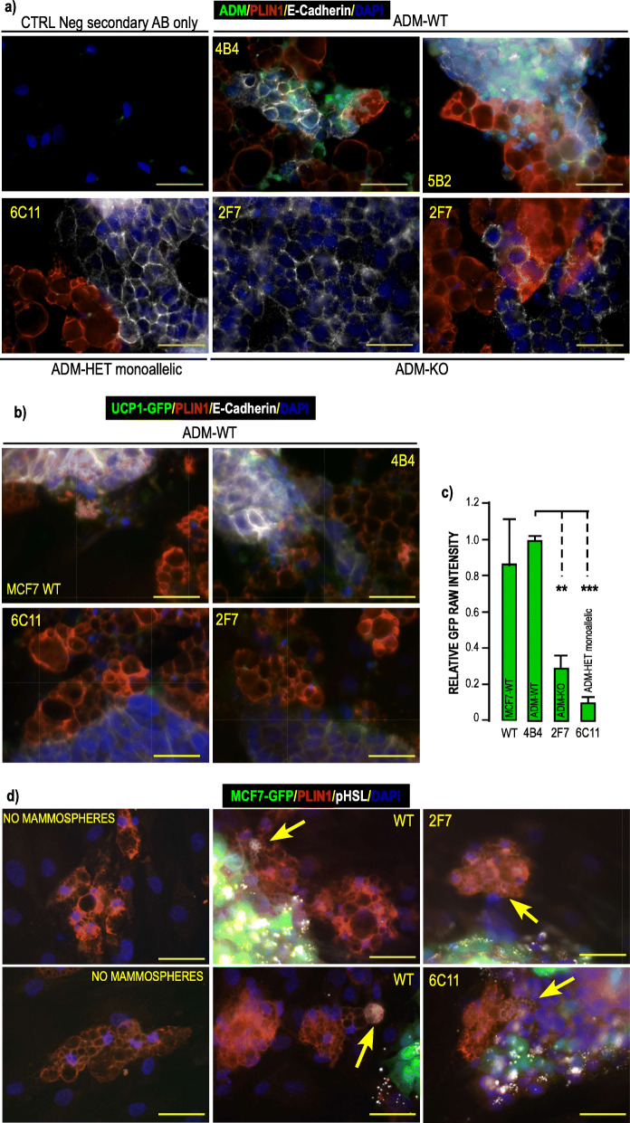
Fig. 6.
Analysis of ADM mutated clones obtained by CRISPR Cas9 technology. a ADM expression in mutated clones obtained by CRISPR Cas9 technology. WT MCF7, ADM-Het and ADM-KO MCF7 cells were grown as mammospheres for 7 days. They were co-cultured on a monolayer of hMADS cells that had been differentiated for 14 days. Co culture of the 2 cell types lasted for 4 days. ADM (green) PLIN1 (red) and E-Cadherin (white) expressions were visualized using specific antibodies. DAPI was used to label the nuclei (Blue). Images were recorded using the very same settings for ADM signal (i.e. 300 m seconds.). Magnification 40X, scale bar 50 μM. b ADM mutated clones are less efficient at inducing UCP1 expression in hMADS- adipocytes. WT-MCF7, ADM-Het and ADM-KO MCF7 cells were grown as mammospheres for 7 days. They were co-cultured for 4 days on a monolayer of hMADS cells expressing GFP under control of the UCP1 promoter that had been differentiated for 14 days. PLIN1 (red) and E-Cadherin (white) expressions were visualized using specific antibodies. GFP expression was visualized in green. DAPI was used to label the nuclei (Blue). Images were recorded using the very same settings. Magnification 40X, scale bar 50 μM. GFP signals were quantified using ImageJ software and compared to signals obtained in the presence of the WT MCF7 cells. c Quantification of the signals. Means were calculated from 3 independent experiments performed in duplicate on at least 3 distinct recordings for each coverslip. The fluorescence signal measured as “raw integrated density” and compared to the signal obtained with WT-MCF7 cells. (**p < 0.01, ***p < 0.001). d) ADM mutated clones induced HSL phosphorylation in hMADS- adipocytes. WT MCF7, ADM-Het and ADM-KO MCF7 expressing GFP cells were grown as mammospheres for 7 days. They were co-cultured for 3 days on a monolayer of breast adipocytes that had been differentiated for 21 days. PLIN1 (red) and pHSL (white) expressions were visualized using specific antibodies. GFP expression was visualized in green. DAPI was used to label the nuclei (Blue). Images were recorded using the very same settings. Arrows indicate pHSL labelling in cells expressing PLIN1. Magnification 40X, scale bar 50 μM. Single channels pictures are presented in supplementary figure 8

