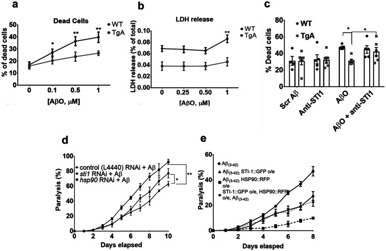
Fig. 1.
Overexpression of STI1 protects both C. elegans and primary hippocampal mouse neurons from Aβ-toxicity. a The average percent of dead cells ((# of dead cells)/(# of live + # of dead cells))× 100) in E17.5 primary hippocampal neuronal cultures from WT or TgA embryos. Cells were imaged in 4 well dishes from 8 random fields (N = 6 individual embryos/genotype). Dishes were treated with either no Aβ, 0.1, 0.5 or 1 μM AβOs for 48 h. b Likewise, WT or TgA primary hippocampal neurons were treated with 0, 0.25, 0.5 or 1 μM for 48 h, and LDH release was measured using colorimetric assay, read at 450 nm. N = 3–4 individual embryos for WT hippocampal cultures for each condition and N = 5 individual embryos per condition for TgA hippocampal cultures. c Quantification of percentage of cell death in hippocampal neuronal cultures treated for 48 h with 1 μM scrambled Aβ control, antibody against STI1 (1:500), AβO alone (1 μM) or dishes treated with both AβO (1 μM) and anti-STI1 (1:500). At least four individual embryos were used for each condition and genotype. d Percentage of body paralysis over 10 days in nematodes expressing Aβ(3–42) (strain CL2006 (dvIs2 [pcL12(unc-54/human Aβ peptide minigene) + pRF4]) in the bodywall muscle and treated with empty vector control RNAi (black triangle), sti-1 RNAi (black circle), or hsp-90 RNAi (black square). e Percentage of paralysis in worms expressing Aβ(3–42) (black circle), Aβ(3–42) worms overexpressing HSP90 in body wall (strain AM988 (rmIs347(unc-54p::HSP-90::RFP)) (black square), Aβ(3–42) worms overexpressing STI-1 in muscle cells (strain PVH40 (PPI1972 (unc-54p::STI-1::GFP);dvIs2))) (black triangle) and Aβ(3–42) worms overexpressing both STI-1 and HSP-90 in the bodywall muscle (strain PVH71 (rmIs347(unc-54p::HSP-90::RFP);(unc-54p::STI-1::GFP);dvIs2) (black stars). For C. elegans experiments, 100 age synchronized animals were used for analyses. For panels a and b, data were analyzed using Two-Way ANOVA, with Sidak’s or Bonferroni’s post-hoc tests for multiple comparisons, respectively, comparing WT vs TgA across the different concentrations of the AβOs. For both panels c and d, groups were analyzed using Wilcoxon statistics, comparing to Aβ(3–42) expressing organisms. *p < 0.05, ** p < 0.01. All data are mean ± SEM. Raw data available for Figure 1: 10.6084/m9.figshare.12115614

