Fig. 3.
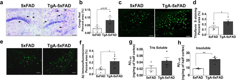
Increased amyloidosis in young male 5xFAD mice overexpressing Hsp90 co-chaperone STI1. a Representative brightfield micrographs of Congo Red labelling in the CA1/Subiculum, at 10X magnification and b quantification of average area fraction (% area) of Congo Red staining across the whole hippocampus in 3–5-month-old male 5xFAD and TgA-5xFAD mice. Black arrows indicate plaques labelled with Congo Red. On each graph, black circles represent individual 5xFAD mice and black squares are individual TgA-5xFAD mice. c Thioflavin-S staining representative images from CA1/Subiculum hippocampal subfield and d, quantification of average percent area of the whole hippocampus with Thioflavin S staining. e Aβ (6E10 antibody, Biolegend) immunoreactivity in CA1/Subiculum and f quantification of average percent area across the whole hippocampus with Aβ immunofluorescence. Concentration (ng/mg) of Tris Soluble g, and GHCl Insoluble h, Human Aβ1–42 in cortex (1 hemisphere) from 3 to 5-month old 5xFAD and TgA-5xFAD male mice. All scale bars = 25 μm. Data are presented as mean ± SEM, and experiments were analyzed using unpaired t-test. *p < 0.05, ** p < 0.01. Raw data available at: 10.6084/m9.figshare.12115713

