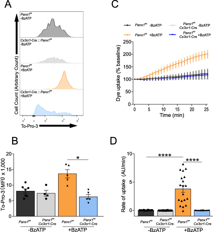
Fig. 2.
Panx1 activity is reduced in myeloid cells (EGFP positive) from Cx3cr1-Cre::Panx1fl/fl mice compared to the Panx1fl/fl mice. BzATP-induced To-Pro 3 uptake via Panx1 activation was assessed using flow cytometry (a and b) and confocal microscopy (c and d). At 3-days post-injury, the ipsilateral cortex was isolated and dissociated to obtain Cx3Cr1+ cells. a Representative example of flow cytometry analysis for each group showing To-Pro 3 uptake. b Quantification of mean fluorescence intensity of the different groups (n = 4 or 6 mice per group; One-way ANOVA (F3,17 = 11.56), Tukey’s post hoc test). c Kinetic of To-pro-3 uptake in isolated Cx3Cr1+ cells measured by confocal microscopy. d Rate of To-pro-3 uptake. At least 20 cells from three separate experiments were used to obtain the rate. One-way ANOVA (F3,96 = 48.87) and Tukey’s post hoc test. *P > 0.05, **** P < 0.0001

