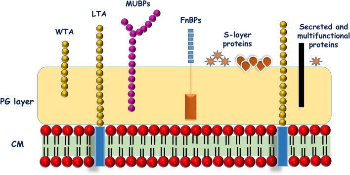
Fig. 5.
Diagrammatic representation of various cell surface-associated components of lactic acid bacteria. (CM, Cell membrane; PG, Peptidoglycan; WTA, Wall teichoic acids; LTA, Lipoteichoic acid; MUBPs, Mucin binding proteins), FnBPs, Fibronectin binding proteins; S-layer, Surface layer)(CM, Cell membrane; PG, Peptidoglycan; WTA, Wall teichoic acids; LTA, Lipoteichoic acid; MUBPs, Mucin binding proteins), FnBPs, Fibronectin binding proteins; S-layer, Surface layer)

