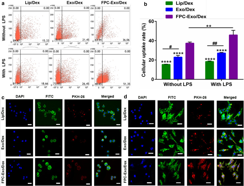
Fig. 4.
Uptake of Lip/Dex, Exo/Dex and FPC-Exo/Dex by RAW264.7 with or without LPS activation. a Representative images of Flow cytometry analysis showing uptake of Dex preparations labeled by PKH 67 in RAW264.7 cells with or without LPS activation. b Uptake rate of different Dex preparations by flow cytometry (n = 3, **p < 0.01, ****p < 0.0001 vs cells treated with FPC-Exo/Dex, #p < 0.05, ##p < 0.01 are cells treated with Lip/Dex compared to Exo/Dex). c Confocal microscopy showing uptake of different Dex preparations labeled by PKH 67 in RAW264.7 cells without LPS activation. Dex preparations appear in red; nucleus, blue; and cytoplasm, green. Scale bar, 20 μm. d Confocal microscopy showing uptake of different Dex preparations labeled by PKH 67 in RAW264.7 cells without LPS activation. Dex preparations appear in red; nucleus, blue; and cytoplasm, green. Scale bar, 20 μm

