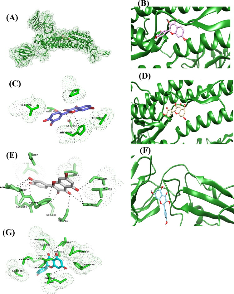
Figure 1.
The putative binding site of fisetin, quercetin and kamferol on SARS-CoV-2S protein. A) The cartoon showing the structure and surface of SARS-CoV-2 S protein, chain A and the binding site of fisetin, quercetin and kamferol, on it. Fisetin, quercetin and kamferol are shown in ball and stick model in purple, pale orange and cyan colour respectively. B) Binding site of fisetin on SARS-CoV-2 S protein. C) The residues interacting with the fisetin. D) Binding site of quercetin on SARS-CoV-2 S protein. E) The residues interacting with the quercetin. F) Binding site of kamferol on SARS-CoV-2 S protein. G) The residues interacting with the kamferol.

