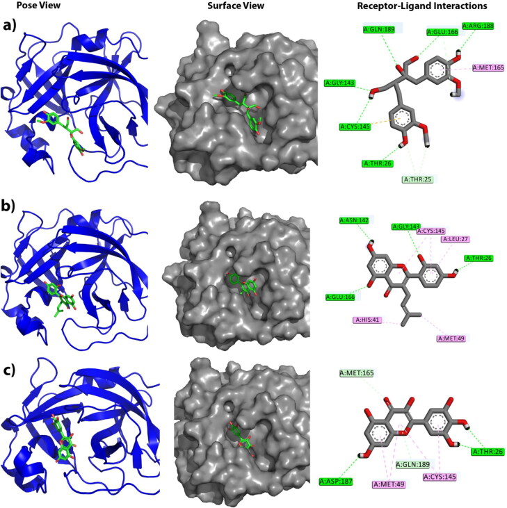
Figure 1.
The figure illustrates different binding modes of selected compounds within the active and catalytic sites of main protease. The alphabetical orders indicate the respective complex of alpha-ketoamide, Carinol, Albanin and Myricetin, respectively. The block and line colors at receptor-ligand interactions such as green, light sky and pink define conventional hydrogen bonding, C-H bonding and hydrophobic interactions, respectively.

