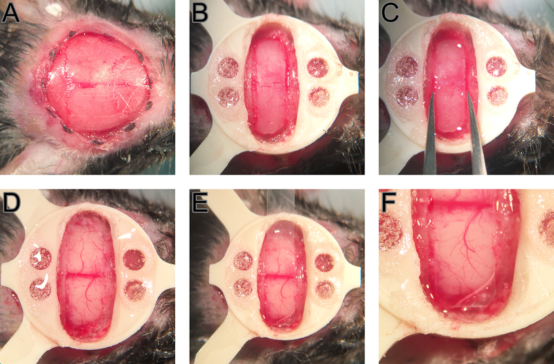
Figure 1.
Surgical steps. A crosshatch pattern is carved on the skull anterior to the bregma and posterior to the lambda (A). A PEEK head plate is attached and craniotomy borders are drilled (B). Bone flap is “loosened” by grabbing form the long edges and repetitively moving anterior and posteriorly (C). Minor dural bleedings are controlled using sterile saline and surgifoam (D). PMP is positioned to fit inside the craniotomy fully leaving minimum gap between the brain and the polymer (E and F).

